To explore abnormalities and to understand the mechanisms underlying functional gastrointestinal disorders and brain-gut axis, we record gastrointestinal motility in anesthetized and freely-moving conscious rodents.
In our lab, we fabricate and tailor our own strain gauge transducers for distinct recording approaches.
Fig.1 represents details of gastric motility recording from an anesthetized rat. A: a rat fixed on a stereotaxic frame, B: brain stem is exposed by blunt dissection, C: a miniature strain gage transducer placed on the proximal stomach, D: a glass micropipette is positioned in caudal medulla targeting left dorsal motor nucleus of the vagal nerve, E: enhanced gastric motility in following microinjection of thyrotropin releasing hormone (TRH). P: indicates the tip of the pipette.
Fig.2 includes the details of gastric motility recording from freely-moving conscious rats. A: a strain gauge transducer specifically fabricated for gastrointestinal motility recording in awake rats, B: serosal implantation of transducer onto the distal stomach, C&D: the recording setup and simultaneous recording of gastroduodenal motility in two awake rats, E: a representative trace of fasting motor pattern recorded from duodenum. Triangles depict the phase-III-like contractions of migrating motor complex (MMC).
We record gastric motility not only for investigation of efferent visceromotor functions, it is also used for evaluation of the vago-vagal afferent gastrointestinal reflexes.
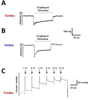
Esophageal Gastric Relaxation (EGR) reflex refers to the muscular relaxation of the proximal stomach in response to existence of food in the lower esophagus. The food-induced afferent sensory signals are transmitted to the brain via the afferent branch of the vagus nerve which is followed by a gastric relaxation triggered by efferent motor signals from dorsal nucleus of the vagus nerve (DMV). Fig.3 represents the summary of the motor and sensory pathways contribute to the regulation of EGR (Modified from study of Hermann and colleagues; Am J Physiol Regul Integr Comp Physiol. 2006; 290 (6): R1570-1576).
Similarly, gastric accommodation reflex (GAR) is a vagal nerve mediated reflex, which is associated with reduction in gastric tone along with an increase in gastric volume and gastric compliance. Below is the summary of the motor and sensory pathways that regulate GAR (modified from Barrett, K. E. Gastrointestinal physiology, 2nd Ed.; 2006).

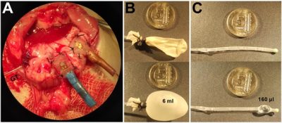
The transducers fixed on gastric fundus and corpus (A) and the custom-made balloon catheters for evaluation of GAR (B) and EGR (C)
The figure below represents examples of our EGR and GAR recordings performed in rats.
Son güncelleme : 9.10.2023 00:29:57
