Immunohistochemistry
Immunohistochemistry (IHC) detects the antigens in cells within a tissue section, where antibody-antigen binding can be visualized by color-producing reactions through specific enzymes including horseradish peroxidase and alkaline phosphatase. On the other hand, in immunofluorescence (IF) method to visualize an antigen-antibody binding , a fluorescent dye is used such as fluorescein isothiocyanate (FITC), tetraethylrhodamine isothiocyanate (TRITC).
In our lab, we perform immunohistochemical staining on wide variety of samples including paraffin, cryo- and freezing sections obtained from brain and gut, as well as cultured neuronal cell lines.
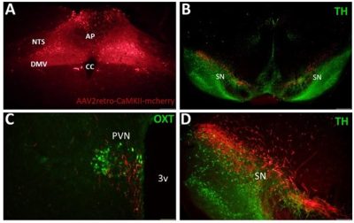
Fig.1Fluorescence immunoreactions in several brain sections harvested 28 days after AAV vector application in dorsal motor nucleus of the vagal nerve (A), B: tyrosine hydroxylase-positive cells substantia nigra (green) C: oxytocin-positive cells (green) in paraventricular nucleus of hypothalamus, D: closer view of substantia nigra. Red: the neuronal cells transfected with AAV2retro-CaMII-mcherry. AP: area postrema, DMV: dorsal motor nucleus of the vagal nerve, NTS: nucleus of the solitary tract, CC: canalis centralis, PVN: hypothalamic paraventricular nucleus, SN: substantia nigra, 3v: third ventricle, TH: tyrosine hydroxylase, OXT: oxytocin.
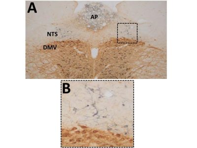
Fig.2 A: The cells expressing chat choline acetyltransferase (ChAT; brown) and tyrosine hydroxylase (TH; black) in the rat dorsal vagal complex, B: the closer view of the boxed area. AP: area postrema, DMV: dorsal motor nucleus of the vagal nerve, NTS: nucleus of the solitary tract.

Fig.4 The tyrosine hydroxylase (TH)-positive cells in rat substantia nigra, and ventral tegmental area 7 days after a unilateral application of 6-OHDA (left). Note that the TH-positive cells are fully preserved in the contralateral hemisphere (right).
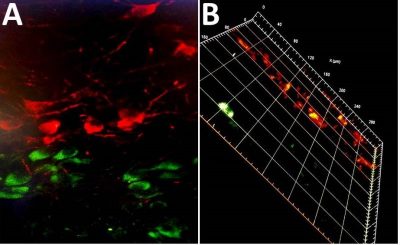
Fig.3Confocal microscopy of the cells expressing chat choline acetyltransferase (ChAT; green) and tyrosine hydroxylase (TH; red) in the rat dorsal vagal complex (A), and z-sectioning image (B).
Son güncelleme : 9.10.2023 00:40:41