In-vivo brain microdialysis is a minimally-invasive sampling technique that is used for continuous measurement of released compounds into the extracellular fluid of the central nervous system through a semi-permeable membrane. Recovered organic analytes in a dialysate include, but not limited to neurotransmitters, neuropeptides, and cytokines. The structure of a microdialysis probe is shown in Figure.1.
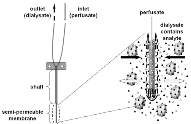
Figure.1: Schematic illustration of a microdialysis probe.
Depending on the targeted region in brain, we use a variety of probes with different membrane and shaft length combinations. Generally, water-soluble compounds diffuse across the microdialysis membrane, while their recovery gradually decreases as the molecular weight of the analyte exceeds 25% of the probe membrane cutoff.
We use microdialysis technique in;
-
Freely-moving conscious rodents and,
-
Anesthetized rodents that are placed in a stereotaxic frame.
Brain microdialysis technique in freely-moving animals enables us to perform multi session sampling for monitoring chronic changes. On the other hand, doing so in anesthetized animals is useful for investigation of the acute changes occurred in response to a specific stimulus such as microinjection of a drug, stress loading or visceral distension.
For this purpose, rodents undergo stereotaxic microdialysis surgery, as shown in Figure.2.
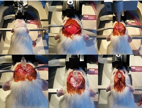
Figure.2: Stereotaxic implantation of a guide cannula into the right hypothalamic paraventricular nucleus in a rat.
Followed by a 7-day recovery period, microdialysis sampling is performed in freely-moving conscious rodents, as seen in Figure.3.
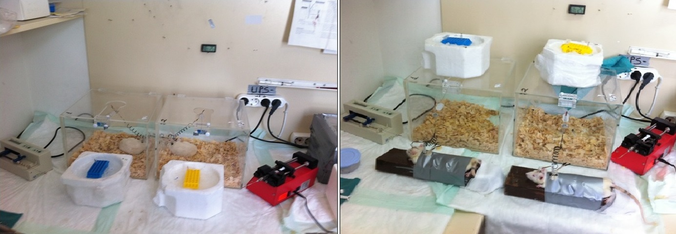
Figure.3: Following collection of the basal microdialysates (left), rats are loaded with restraint stress (right) and microdialysis sampling is continued during stress.
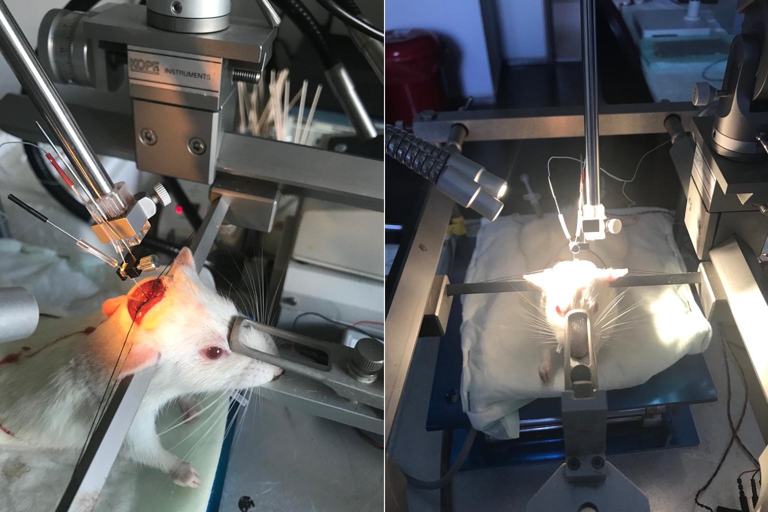
Figure.4: Microdialysis sampling from dorsal vagal complex in an anesthetized rat placed on in a stereotaxic frame.
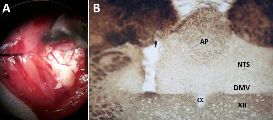
Figure.5: A microdialysis probe that is placed in left dorsal vagal complex (A), the post-mortem histological verification of the probe placement in left dorsal vagal complex (B).
Son güncelleme : 4.03.2024 00:02:03
