The blood-brain barrier is a highly selective semipermeable diffusion barrier which circumvents the influx of most therapeutics into the brain. This barrier is crossed by icv injection, allowing direct delivery of drugs and chemicals to the rat central nervous system. Generally, we perform icv applications with two approaches:
-
a single, acute administration, and
-
repeated, chronic administrations
Neurotoxins, drugs, antibodies, viral vectors, and stem cell preparation can be directly deposited into the lateral ventricle; whereas, a custom-made port (icv cannula) is placed into the ventricle for easy access.

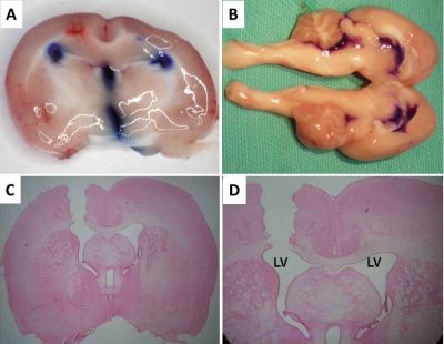
In rats, we usually prefer left/or lateral ventricle for icv applications. The specific coordiantes for are obtained from the reliable Rat Brain Atlas of Paxinos. In reference to the Bregma (Fig1.A), we usually pick -1.2 mm as the AP cooordinate (Fig.1B). LV depicts right lateral ventricle.
Cannulation Procedure

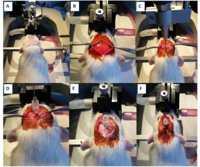
An anesthetized rat is placed in a stereotaxic frame (A). According to appropriate coordinates we create a burr hole using a dental drill attached on a stereotaxic manipulator (B-C). After the cannula is placed into the lateral ventricle, it is fixed onto the skull surface with the aid of an anchor screw and dental cement (D-E). Then the incision is closed with silk suture (F).
Generally; following icv cannulation, an operated rat is housed individually in the home cage for 5 days for recovery.
Functional Verification
After the recovery, to verify the cannula placement in the lateral ventricle, 100 ng angiotensin II is administered through the icv cannula and rat is then returned to home cage with access to a water bottle (A). The latency to drink is recorded so that the animals failed to drink within 120 sec are excluded from upcoming experiments (B).

Morphological Verification

Once the experiments are done, a proper cannula placement is also verified by icv injection of 5-10 µl of methylene blue. Following the application, brain is cut sagittally, then the spread of methylene blue in ventricles is examined (A-B). The coronal sections can also be examined under a light microscope after rapid fast red staining (C-D).
Eklenme tarihi :9.10.2023 00:10:11
Son güncelleme : 4.03.2024 00:03:34
Son güncelleme : 4.03.2024 00:03:34
